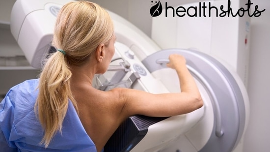
A report that says everything looks normal can bring a sense of relief. But when it comes to breast health, does a normal mammogram always tell the whole story? Not necessarily. Mammography uses X-rays to examine breast tissue and can detect cancer early, sometimes before a lump can be felt. However, it may not detect every cancer, and its accuracy can vary from woman to woman, particularly depending on breast density. Understanding these limitations helps women make informed decisions about screening and recognise when further medical evaluation may be needed.
Awareness is particularly important in India
The Ministry of Health and Family Welfare projected 2,40,381 breast cancer cases in the country in 2025. Early detection can make a meaningful difference to breast cancer outcomes. "The World Health Organisation (WHO) reports that the five-year survival rate for breast cancer exceeds 90% in high-income countries, compared with 66% in India. This gap highlights the challenges many women face in accessing timely diagnosis and treatment," Medical oncologist Dr Yashwant Kashyap (TMH Mumbai), MD General Medicine, MBBS (BHU Varanasi), tells Health Shots. Greater awareness, appropriate screening, recognising changes in the breast and seeking medical advice promptly can help women receive the care they need without delay.
Why may a normal report not tell the whole story?
Mammography plays an important role in breast cancer screening. Still, its ability to detect cancer can vary from woman to woman. In contrast, diagnostic mammograms help investigate a breast change or an abnormal screening result. One factor that can affect how clearly a mammogram reveals an abnormality is breast density.
Dense breasts contain more glandular and connective tissue relative to fatty tissue. Since both dense tissue and some cancers appear white on a mammogram, cancer can sometimes be obscured by the surrounding tissue. This is particularly relevant in India, where research has highlighted breast density alongside gaps in screening infrastructure and trained personnel as challenges to early detection. Mammography sensitivity (the ability to identify cancer when it is present) has been reported to range from 64% to 90%. This means that, although mammography is a valuable screening tool, a normal result cannot rule out cancer in every case.
What do normal and abnormal results mean?
When a mammogram appears normal despite cancer being present, it is called a false-negative result. The American Cancer Society estimates that screening mammograms miss about one in eight breast cancers, with false negatives more likely in women with dense breasts. This is why a new lump, skin dimpling, nipple inversion, unusual discharge or persistent breast change should be medically assessed, even after a recent normal mammogram.
- The reverse can also happen. A false-positive result occurs when a mammogram looks abnormal but further evaluation finds no cancer. Such findings can cause worry and lead to additional appointments, tests and expense. However, an abnormal report is not a cancer diagnosis; it means the finding needs closer examination.
- Depending on the findings, the doctor may recommend a diagnostic mammogram for focused X-ray images, an ultrasound to assess a lump, an MRI for women at higher risk or in selected cases, or a biopsy to examine a tissue sample and confirm whether cancer is present.
- Advances in screening technology may also help improve detection. Three-dimensional (3D) mammography, also called digital breast tomosynthesis, creates images of thin layers of breast tissue. This may improve detection of some cancers and reduce callbacks in certain women. However, availability and cost can be limiting factors.
What should women in India know about screening?
{{/usCountry}}When a mammogram appears normal despite cancer being present, it is called a false-negative result. The American Cancer Society estimates that screening mammograms miss about one in eight breast cancers, with false negatives more likely in women with dense breasts. This is why a new lump, skin dimpling, nipple inversion, unusual discharge or persistent breast change should be medically assessed, even after a recent normal mammogram.
- The reverse can also happen. A false-positive result occurs when a mammogram looks abnormal but further evaluation finds no cancer. Such findings can cause worry and lead to additional appointments, tests and expense. However, an abnormal report is not a cancer diagnosis; it means the finding needs closer examination.
- Depending on the findings, the doctor may recommend a diagnostic mammogram for focused X-ray images, an ultrasound to assess a lump, an MRI for women at higher risk or in selected cases, or a biopsy to examine a tissue sample and confirm whether cancer is present.
- Advances in screening technology may also help improve detection. Three-dimensional (3D) mammography, also called digital breast tomosynthesis, creates images of thin layers of breast tissue. This may improve detection of some cancers and reduce callbacks in certain women. However, availability and cost can be limiting factors.
What should women in India know about screening?
{{/usCountry}}Screening should be guided by a woman’s age, medical history and individual risk. According to the Federation of Obstetric and Gynaecological Societies of India (FOGSI), routine population-based mammography is generally not recommended for women under 40, as breast cancer incidence is lower and denser breast tissue can reduce mammography sensitivity. Instead, the focus should be on breast self-awareness, clinical breast examination and targeted imaging for women at higher risk.
- As women get older, the approach to screening can change. For women aged 40–49, screening decisions should be individualised. For those aged 50–69, screening should be every two years. In contrast, for women aged 70 and above, screening should consider life expectancy, other health conditions, and screening history.
- Age alone, however, does not determine a woman’s screening needs. Those with a strong family history of breast cancer, known BRCA1 or BRCA2 mutations, or other significant risk factors should consult a healthcare professional to assess their risk and determine whether earlier or more intensive screening is appropriate.
- While mammography remains an important tool for early detection, understanding individual risk, choosing appropriate screening options and seeking medical advice for any new or persistent breast changes, even after a normal mammogram, are essential steps towards timely diagnosis.
(Disclaimer: This article is for informational purposes only and is not a substitute for professional medical advice. Always seek the advice of your doctor with any questions about a medical condition.)
Catch your daily dose of Fashion, Taylor Swift, Health, Festivals, Travel, Relationship, Recipe and all the other Latest Lifestyle News on Hindustan Times Website and APPs.
Catch your daily dose of Fashion, Taylor Swift, Health, Festivals, Travel, Relationship, Recipe and all the other Latest Lifestyle News on Hindustan Times Website and APPs.