In explaining the significance of the research that won this year’s prize for Physiology or Medicine on Monday, the Nobel Committee begins by quoting from a letter Virginia Woolf wrote to a friend in 1932: “My own brain is to me the most unaccountable of machinery — always buzzing, humming, soaring, roaring, diving, and then buried in mud. And why? What’s this passion for?”
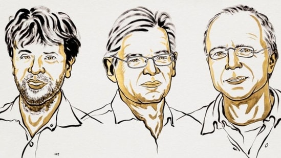
The three laureates who share the Nobel, Karl Deisseroth, Peter Hegemann and Georg Nagel, made discoveries that opened a new technique, called optogenetics, which helps scientists hunt for answers to the questions that puzzled Woolf and billions before her. How does a single organ, which weighs just over a kilogram, hold memories, form feelings, and control the rhythm of breathing and the sleep cycle?
We know different nerve cells control different brain functions, but earlier methods struggled to identify which kinds of cells were responsible for which kinds of functions. Today, optogenetics makes it possible to switch on or switch off the activity of individual nerve cells. This helps unlock the brain’s mysteries, including what happens in the organ leading to disorders ranging from depression and anxiety to Alzheimer’s and Parkinson’s diseases. The long-term goal is to use optogenetics for medical treatment, and there have been breakthroughs in that direction, including partial restoration of vision in people with an eye disease called retinitis pigmentosa.
{{/usCountry}}We know different nerve cells control different brain functions, but earlier methods struggled to identify which kinds of cells were responsible for which kinds of functions. Today, optogenetics makes it possible to switch on or switch off the activity of individual nerve cells. This helps unlock the brain’s mysteries, including what happens in the organ leading to disorders ranging from depression and anxiety to Alzheimer’s and Parkinson’s diseases. The long-term goal is to use optogenetics for medical treatment, and there have been breakthroughs in that direction, including partial restoration of vision in people with an eye disease called retinitis pigmentosa.
{{/usCountry}}The seeds for this were laid more than 30 years ago by Hegemann (currently with Humboldt University of Berlin) and Nagel (University of Würzburg, Germany), and followed up by Deisseroth (Howard Hughes Medical Institute, and Stanford University). The work started with a humble alga.
The protein
The green alga Chlamydomonas has only one cell, but reacts astonishingly to light. When it is put into a petri dish and one side of the dish is illuminated, the alga swims towards it. It senses the light with a tiny orange dot called the eyespot.
Also read: Unknown pneumonia: What Russia's first remarks say and don't say about Siberia plague fears
In the 1990s, Hegemann, then at the Max Planck Institute for Biochemistry in Martinsried, found that the speed of the eyespot’s responses was incredibly fast. It produced an electrical impulse just half a millisecond after light reached it, more than 20 times faster than the corresponding process in the human eye, which takes at least 10 milliseconds.
In the human eye, a complex chain of events is involved. The final step is an ion channel (a specialised type of protein) that allows charged ions to flow, creating an electrical signal. Hegemann suggested that in the alga, a single protein must be not only capturing the light but also acting as the ion channel. The hypothesis was met with scepticism then, but Hegemann, in collaboration with Nagel, went on to identify such a protein.
They used the findings of Japanese researchers, who had mapped the DNA of Chlamydomonas. From the DNA, Hegemann and his colleagues found two genes with promising properties and sent them to Nagel, then at the Max Planck Institute for Biophysics in Frankfurt, who had the expertise necessary to hunt for the protein.
Nagel injected the two genes, separately, into different batches of frog eggs. The eggs began to mass produce two unknown proteins. Nagel found that the proteins were indeed ion channels that opened when exposed to light, proving the hypothesis Hegemann had made a decade earlier.
These proteins were named channelrhodopsin-1 and channelrhodopsin-2. It is the second that opened the road to optogenetics. When Hegemann and Nagel introduced the gene for channelrhodopsin-2 into embryonic human kidney cells, and in kidney cells from hamsters, the cells became light-sensitive and produced an electrical signal when exposed to light.
Deisseroth, at Stanford, then took this work forward to nerve cells, first in a petri dish, and then in living mice.
From petri dishes to mice
Deisseroth had his own research group, searching for a protein that could trigger electrical impulses in nerve cells. When he heard about channelrhodopsin-2, he wrote to Nagel, who sent him the DNA encoding the protein.
When Deisseroth introduced the DNA into rat nerve cells cultured in petri dishes, the cells produced channelrhodopsin-2, and reacted to blue light, producing a nerve signal. His team and other groups soon found more proteins that can turn light-responsive nerve cells on and off. In 2006, this method was given the name optogenetics.
So far, the work had only been done in cultured nerve cells. Then in 2007, Deisseroth activated nerve cells in the brains of living mice.
By introducing the gene for channelrhodopsin-2 into a specific type of mouse nerve cell, and illuminating the nerve cells, Deisseroth and his team could control movements of the mouse whiskers. In another experiment using optogenetics, Deisseroth and other researchers woke sleeping mice in a controlled manner.
Then in 2012, they made a bigger breakthrough. Deisseroth and Susumu Tonegawa (the 1987 Nobel Prize winner) identified which nerve cells in the mouse brain appeared to form a memory of a fearful experience, and later reactivated these nerve cells. The mice showed signs of fear, despite not being in danger at the time.
Why it matters
In a network of billions of human nerve cells, optogenetics gives researchers a tool to hunt and identify which cells and neural circuits are involved in particular brain functions. They have identified neural networks that govern the sense of pain, thirst, and the circadian rhythm, among various functions.
Optogenetics can also be used to map cell functions outside the brain; for example, Deisseroth has shown that heart rhythm can affect emotions such as anxiety, while other researchers have identified gut cells that can explain why some people would rather eat sugar than sweeteners. Optogenetics has also enabled better understanding of the neural activity involved in depression, anxiety, schizophrenia, Alzheimer’s disease and Parkinson’s disease.
As for medical treatment, ongoing clinical trials are attempting to restore vision in people who have become blind due to a disease called retinitis pigmentosa. When a protein similar to channelrhodopsin was inserted in the retina of a blind person, the person regained some vision and, using special glasses, was able to discern and grasp objects on a table.