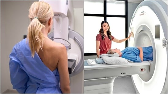
Whenever you go for a breast checkup, your doctor might recommend a mammography, ultrasound, or an MRI interchangeably. But what decides which test is correct for you? All these imaging tests serve different purposes, and the right one depends on factors such as a woman's age, breast density, medical history and symptoms.
In an interaction with HT Lifestyle, Dr Jyoti Arora, principal lead of women’s imaging at Apollo Athena Women’s Cancer Centre, explained the difference between mammography, ultrasound, and MRI.
“‘Why did my doctor ask for an ultrasound when my friend was told to get a mammogram?’ It is a question I hear almost every day. The answer is simple: mammography, ultrasound and MRI are three different ways of looking at the breast, and each sees something the others can miss,” Dr Jyoti explained. According to her, choosing the right one is not about which is newer or costlier; it is about matching the test to the woman and to the problem.
The mammogram: the screening workhorse
A mammogram is a low-dose X-ray of the breast, Dr Jyoti noted. Explaining what a mammogram does, she stated, “Its great strength is that it can detect tiny calcium deposits, called microcalcifications, which are sometimes the earliest sign of cancer, long before any lump can be felt.”
{{/usCountry}}A mammogram is a low-dose X-ray of the breast, Dr Jyoti noted. Explaining what a mammogram does, she stated, “Its great strength is that it can detect tiny calcium deposits, called microcalcifications, which are sometimes the earliest sign of cancer, long before any lump can be felt.”
{{/usCountry}}“Large studies across several countries have shown that regular screening mammography lowers deaths from breast cancer, especially in women aged 40 to 74. This is why it remains the standard test for screening, meaning checking healthy women who have no complaints,” she added.
However, Dr Jyoti stressed that its limitation is density. “Breasts are made of fatty tissue and dense tissue (glands and fibrous tissue). On a mammogram, fat appears dark, while dense tissue and most cancers appear white. A cancer hiding in dense tissue can be like a snowball in a snowstorm. Many Indian women, particularly younger ones, have dense breasts, which is why mammography alone is not always enough. A newer version, 3D mammography (tomosynthesis), takes images in thin slices and helps reduce this masking effect,” she explained.
Ultrasound: the problem-solver
Ultrasound uses sound waves, which carry no radiation, so it is safe during pregnancy and for young women, Dr Jyoti noted. “Its special skill is telling fluid from solid. A smooth, fluid-filled cyst, which is usually harmless, looks very different from a solid lump that needs a closer look. This is why ultrasound is often the first test for a woman under 30 or 40 who finds a lump, and why it is used alongside a mammogram when the breasts are dense. It also guides the needle when a small sample (biopsy) is taken,” she explained.
Highlighting its limits, Dr Jyoti shared, “It is not a good screening tool by itself, because it can miss microcalcifications, and its results depend heavily on the skill of the person performing it. It can also flag many harmless findings, which may cause worry and extra tests.”
MRI: the detailed map
Lastly, Dr Jyoti shared that magnetic resonance imaging (MRI) uses a strong magnet and radio waves, again with no radiation. Explaining how MRI works, she stated, “After an injection of contrast dye, it shows how blood flows within the breast. Cancers tend to grow their own abnormal blood vessels, so they light up on MRI. It is the most sensitive of the three tests.”
This makes MRI valuable in specific situations, according to Dr Jyoti, who added, “These include women at very high risk, such as those who carry BRCA gene changes or have a strong family history, and women already diagnosed with cancer, where MRI can show the full extent of the disease, check the other breast, and help the surgeon plan. It is also used when other tests are unclear. MRI can even guide a biopsy of an area that no other scan can see clearly.”
However, the trade-off is that MRI can pick up findings that turn out to be harmless, so it is not used to screen every woman. “It is also costlier, takes longer, and is not suitable for everyone, for example, those with certain implants or severe kidney disease.”
How doctors choose
Dr Jyoti advised to see MRI, ultrasound, and mamogram as three tools in a toolbox. As for how doctors asses which tool to use, she explained, “In practice, a woman over 40 with no symptoms will usually begin with a mammogram. A 28-year-old with a lump will usually begin with an ultrasound. A woman with a BRCA mutation may need yearly mammograms and MRI. A woman newly diagnosed with cancer may need all three, so that her surgeon knows exactly what to treat and can often save the breast.”
What you can do
Dr Jyoti advised, “Do not choose the test yourself, and do not be alarmed if your doctor orders a different test from your friend’s. Know your family history and tell your doctor about it. Ask whether your breasts are dense. Above all, do not ignore a new lump, nipple discharge or skin change just because a recent scan was normal. No single test finds everything, and what you feel matters.”
The best imaging is not the most advanced machine; it is the right test, performed well and read by an experienced eye, at the right time.
Note to readers: This article is for informational purposes only and not a substitute for professional medical advice. Always seek the advice of your doctor with any questions about a medical condition.
Catch your daily dose of Fashion, Taylor Swift, Health, Festivals, Travel, Relationship, Recipe and all the other Latest Lifestyle News on Hindustan Times Website and APPs.
Catch your daily dose of Fashion, Taylor Swift, Health, Festivals, Travel, Relationship, Recipe and all the other Latest Lifestyle News on Hindustan Times Website and APPs.