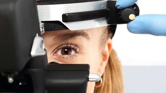
A simple eye examination and retinal imaging test may help improve diagnosis of a progressive neurodegenerative condition called frontotemporal degeneration (FTD). Neurodegenerative diseases in general are challenging to diagnose, and often are confirmed only by direct examination of brain tissue at autopsy.
In a study published online in the journal Neurology, the scientists found that patients with FTD showed thinning of the outer retina — the layers with the photoreceptors through which we see — compared to control participants. The retina is potentially affected by neurodegenerative disorders because it is a projection of the brain.
Prior studies have suggested that patients with Alzheimer’s disease and ALS (amyotrophic lateral sclerosis) may also have thinning of the retina — although a different part of the retina. Thus, imaging the retina may help doctors confirm or rule out frontotemporal degeneration. “Our finding of outer retina thinning in this carefully designed study suggests that specific brain pathologies may be mirrored by specific retinal abnormalities,” said study lead author Benjamin Kim, assistant professor of Ophthalmology at Perelman School of Medicine at the University of Pennsylvania in the US.
The study included a small group of FTD patients and another control group who did not have any neurodegenerative disease. The FTD patients were carefully characterised with clinical examinations, cerebrospinal fluid biomarkers to exclude Alzheimer’s Disease, and genetic testing. The researchers then employed an eye-imaging technology called spectral-domain optical coherence tomography (SD-OCT), which uses a safe light beam to image tissue with micron-level resolution. SD-OCT imaging is inexpensive, non-invasive, and quick.
{{/usCountry}}The study included a small group of FTD patients and another control group who did not have any neurodegenerative disease. The FTD patients were carefully characterised with clinical examinations, cerebrospinal fluid biomarkers to exclude Alzheimer’s Disease, and genetic testing. The researchers then employed an eye-imaging technology called spectral-domain optical coherence tomography (SD-OCT), which uses a safe light beam to image tissue with micron-level resolution. SD-OCT imaging is inexpensive, non-invasive, and quick.
{{/usCountry}}Measurements of the retinal layers of the participants, after adjustments for age, gender, and ethnic background, showed that the outer retinas of the FTD patients were thinner than those in the control participants. This relative thinning of outer retinas was caused by a thinning of two specific portions of the outer retina, the outer nuclear layer (ONL) and ellipsoid zone (EZ). The outer nuclear layer of FTD patients was about 10% thinner than controls, and this ONL thinning was the primary source of the outer retina thinning, the study added.
Follow @htlifeandstyle for more
Catch your daily dose of Fashion, Taylor Swift, Health, Festivals, Travel, Relationship, Recipe and all the other Latest Lifestyle News on Hindustan Times Website and APPs.
Catch your daily dose of Fashion, Taylor Swift, Health, Festivals, Travel, Relationship, Recipe and all the other Latest Lifestyle News on Hindustan Times Website and APPs.