
 E-Paper



Two People Receive the Same Brain Tumour Diagnosis. Their Treatment Paths Differ.
In Bengaluru, brain tumor treatment involves a detailed sequence of diagnosis, surgery, pathology, and rehabilitation. MRI reports guide urgency.

 Prefer HTon Google


A safe outcome needs a sequence that includes diagnosis, surgery, pathology and rehabilitation, not a one-step intervention.'
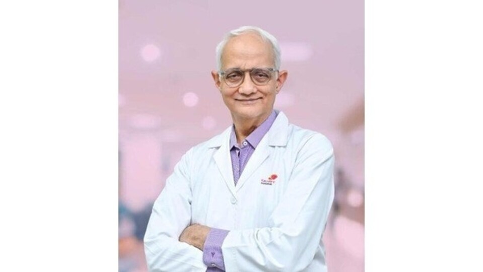
Many families in Bengaluru search for brain tumour treatment in Bangalore and expect one clear recommendation. Some arrive after a seizure at home or at work. Others come with weeks of headache or weakness. An MRI might show a mass with edema and mass effect. These words do not tell the whole story. A safe plan begins with the diagnostic journey and ends with structured recovery.
The diagnostic journey
An MRI report uses phrases that guide urgency and planning. These phrases affect the next steps in your treatment.
Mass lesion or space-occupying lesion -This means an abnormal area taking up space. It is a description. It does not confirm a tumour.
Edema or vasogenic edema - This is swelling around the lesion. Swelling raises pressure inside the skull and often produces headache, nausea or seizures. You might need medicines to control swelling before surgery.
Mass effect - The lesion is pushing on nearby brain structures. Higher mass effect demands faster evaluation.
Midline shift - The brain has shifted from one side to the other. This suggests risk for deterioration and often needs close monitoring in a Neuro ICU.
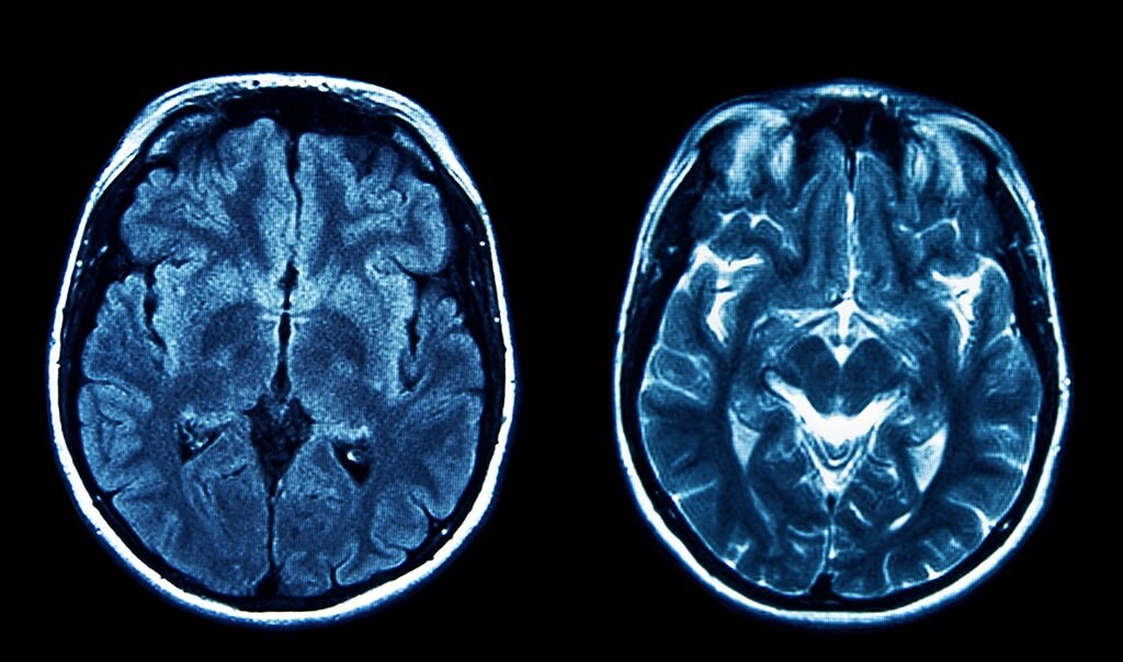
Ring-enhancing lesion - This appearance on contrast MRI fits a wide differential in India. Tumour, tuberculoma or neurocysticercosis are common considerations. Tissue confirmation through biopsy or resection is often necessary.
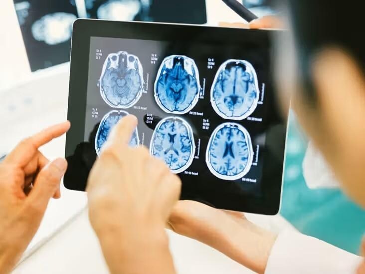
Near eloquent cortex - The lesion sits close to speech, movement or vision areas. This affects how a surgeon plans the corridor and whether mapping or awake surgery is required.
Imaging does not give a final diagnosis. The final diagnosis comes from tissue examination.
Surgical excellence
Brain tumour surgery focuses on maximal safe resection. This means removing as much tumour as possible while protecting speech, movement, memory and personality. The target is safe removal with function preservation. A surgeon uses neuro navigation, high-resolution imaging, a microscope and intraoperative monitoring to map the safest route.
If the tumour is near speech or movement areas, awake mapping helps the team avoid critical networks. During awake surgery, you speak or move your fingers while the team monitors brain function. This reduces the risk of permanent deficits.
Planning includes airway and anaesthesia preparation, seizure prevention, blood management and infection prevention. A coordinated protocol improves safety. Kauvery Hospitals, Bengaluru, follows structured pathways that link preoperative planning, operating room workflow and Neuro ICU readiness.
ICU management after surgery is a critical step. Early complications include bleeding, swelling, electrolyte disturbances, seizures and infection. A dedicated Neuro ICU team monitors neurological status, follows standardised protocols and responds quickly when changes appear.
Pathology drives treatment
An MRI impression does not define the tumour type. A biopsy or surgical specimen provides the final diagnosis. This includes tumour type and grade. Both influence the need for radiation or chemotherapy.
Modern pathology uses integrated reporting based on the World Health Organisation CNS classification. Key molecular markers, such as IDH status and 1p19q codeletion in adult-type diffuse gliomas, shape treatment and follow-up. These markers help teams estimate prognosis and plan adjuvant therapy. They also guide imaging frequency after surgery.
You should ask for a clear explanation of the pathology report. You should know the type, grade and molecular findings. You should know how these details influence the next steps.
When awake, mapping changes risk
Tumours in speech or movement areas need a different plan. Even a small error in these regions can affect daily life. Awake mapping reduces this risk. During the procedure, you perform simple language or motor tasks. The team monitors each response and avoids critical pathways. This increases the chance of preserving speech and movement after surgery.
Rehabilitation
Recovery starts soon after surgery. Neuro rehabilitation includes physiotherapy, speech therapy and cognitive therapy. Early mobilisation improves strength and balance. Speech therapy supports language recovery when tumours affect communication areas. Cognitive therapy helps patients who face attention or memory difficulties after surgery or radiation.
Return to work planning is specific to each patient. Your team should guide you through timelines for light activity, full activity and long-term restrictions. A structured follow-up plan includes seizure control, wound checks, medication review and periodic MRI scans.
Patient decision checklist
When you seek a second opinion or prepare for surgery, bring your scans and reports. Prepare these questions.
- What are the top suspected diagnoses?
- Do you need resection or biopsy first?
- How does tumour location affect speech, movement or vision risk?
- What steps protect function during surgery?
- Is awake mapping recommended?
- What is the ICU plan for the first 24 to 48 hours?
- What details will the pathology report include?
- How will grade and molecular markers influence the next steps?
- What rehabilitation will you need?
- When can you return to work?
Many families ask whether complete removal is possible. Complete removal does not always translate into better outcomes. Safe removal with preserved function matters more. A staged plan based on diagnosis, pathology and recovery provides a better long-term result.
Your care should reflect a sequence. Diagnosis informs surgery. Surgery provides tissue for pathology. Pathology shapes the adjuvant plan. Rehabilitation supports recovery. A centre with coordinated neurosurgery, Neuro ICU, pathology and rehabilitation strengthens each link in this sequence.
A final word from the Brain & Spine Expert of Bangalore
If you have an MRI report that worries you, or if you have been advised brain tumour surgery and want a second opinion, bring your scans, bring your questions, and bring your concerns.
We will walk through the plan together - one step at a time.
For appointments or Expert opinion call 080 6801 6901 or visithttps://www.kauveryhospitalsbangalore.com/
By Dr Ramakrishna Easwaran, Director, Kauvery Institute of Neurosciences, Director of Academics, Kauvery Hospitals Group
Note to readers: This article is part of HT’s paid consumer connect initiative and is independently created by the brand. HT is not responsible for the content’s accuracy or completeness. Readers should verify all information independently.
This content is for informational purposes only. It neither constitutes, nor is a substitute for professional medical advice, diagnosis, or treatment. Always consult a qualified healthcare provider for guidance on your health concerns.
Want to get your story featured as above? click here!


