
 E-Paper



Chandigarh: PGI pioneers minimally invasive brain surgery
Minimally invasive technique for treating giant pineal tumours introduced by Dr Dhandapani SS from Chandigarh’s PGIMER is the first in the world


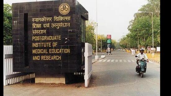
Neurosurgeons of the Post-graduate Institute of Medical Education and Research (PGIMER) have introduced a new method for treating giant pineal tumours using a combination of exoscope and endoscope. This technique, which has just been published in the journal “World Neurosurgery”, is the first of its kind in medical literature.
The pineal gland, which controls our day-night cycle, lies deep inside the brain, making operating on it challenging. Large tumours in this area are especially tricky because they attach to important veins, often requiring a big opening the skull and brain retraction, which increases risks. Despite these techniques, it is hard to locate and separate the tumour from critical veins using traditional methods.
Minimally invasive technique for treating giant pineal tumours introduced by Dr Dhandapani SS of PGIMER’s neurosciences department is the first in the world. Used in over 20 cases with successful results, this method does not incur extra costs for patients as it relies on the institute’s existing equipment.
Dr Dhandapani SS and Dr Chandrasekhar have utilised a novel combination of 3D exoscope, angled endoscope and navigation through a keyhole craniotomy to successfully remove a giant tumour of the pineal gland in a 16-year-old patient.
Used in the initial part of surgery, the 3D exoscope offers greater depth of focus and field of view as compared to traditional microscopic surgery. Once some space became available, an angled endoscope was used to separate the tumour from all critical structures, as the endoscope provides a better panoramic view of corners and crevices.
The doctors have also previously published writings in “Neurology India” on the use of angled endoscopes for pineal region in 2020, and another novel endoscopic technique to get safe biopsy of pineal tumours in “Child’s Nervous System” in 2022.


