
 E-Paper



The inside job: 50 years on, the incredible story of the MRI

 Prefer HTon Google


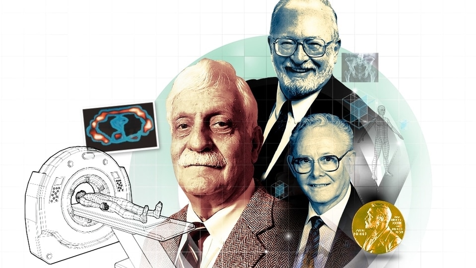
The magnetic resonance image has answered key questions, and is breaching new frontiers. But the road to it was paved with intrigue, luck and loss. Take a look.
It’s a tale of unlikely drama — forgotten heroes, sudden breakthroughs, and through it all, a sustained effort to see more clearly into the human body.
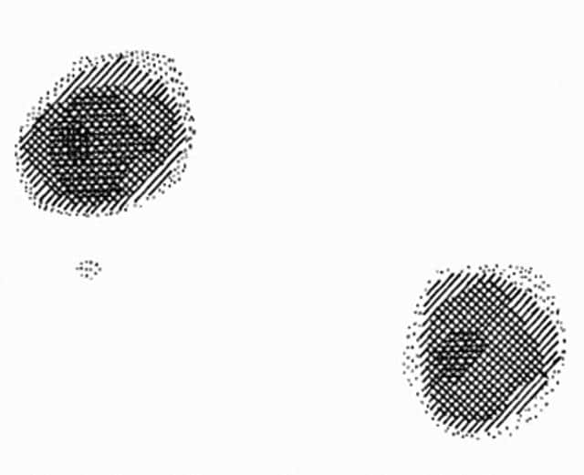
Fifty years ago, in 1973, the first MRI or magnetic resonance image was taken by American chemist Paul Lauterbur of Stony Brook University. In a paper published in the journal Nature, he explained that magnetic resonance could be used to generate an image of a capsule of regular water dunked in a beaker of heavy water (for those who’ve always wondered, heavy water is a form of the liquid in which the hydrogen atoms are all deuterium, a heavy isotope of hydrogen).
Appended to his report was a crosshatched image that showed two circular patches where the test tubes of regular water sat.
Given that 60% of the human body is water in different concentrations, he argued, a magnetic resonance imaging could be used to generate images of the inside of the body. His focus then was on finding a non-invasive way to detect cancerous tumours, because at the time it was almost impossible to see what was going on within muscle and tissue, without slicing into it for a direct view.
X-ray radiation, discovered in 1895, could capture some muscle and bone. The ultrasound scanner, which came into use shortly after World War 2, could image a few internal organs. The Computed Tomography or CT scan, invented in 1971, used a series of X-rays for a cross-sectional view of soft tissue and blood vessels, but none offered a view as clear or complete as the MRI. In addition, the MRI did this without using harmful ionising radiation.
Lauterbur called his imaging technique zeugmatography (from the Ancient Greek for “yoked together”). The name didn’t stick, but the concept would sweep the world, evolving fast as technology improved.
The first MRI of a human body was captured in 1977, in a machine that would be unrecognisable today — it involved, for instance, a large metal grille that locked around the patient’s torso. This machine took five hours to generate a single image, showing a cross-section of the human chest comprising the heart and lungs.
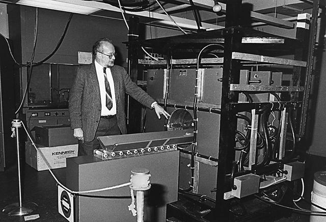
A decade later, the machines would start showing up in hospitals around the world.
The magnetic resonance image is now the definitive tool for detecting nascent tumours, blood clots and a heightened risk of stroke, changes in brain function, tissues, organs and blood vessels.
For kicking off the MRI effort, Lauterbur would eventually win a Nobel Prize, in 2003. But through his life (he died in 2007, aged 77) he was modest about his contribution. He persisted with the idea of this machine, he said in his Nobel lecture, “not because I foresaw all of the medical applications that would follow, but because of the physical uniqueness of the concept.”
***
To be fair, Lauterbur did have reason to be modest. His paper, and the concept it outlined, stood on the shoulders of giant scientific discoveries that came before.
Perhaps most vital among these was nuclear magnetic resonance or NMR, a phenomenon first measured and described 35 years earlier, in 1938, by Columbia University physicist Isidor Rabi.
Rabi was studying the magnetic properties of the nucleus of an atom, and found that, in different substances, atomic nuclei, which are tiny magnets themselves, respond or “resonate” differently to external magnetic fields. This nuclear magnetic resonance, he realised, could be used to study the chemical compositions of different substances. It was a discovery for which he won the Nobel Prize in Physics, in 1944.
In 1946, physicists Felix Bloch and Edward Mills Purcell independently demonstrated NMR spectroscopy for the first time on liquids and solids, and developed precise measurements for this reaction in different materials. This won them the Nobel Prize in Physics, in 1952.
In 1966, Swiss physical chemist Richard Ernst found that NMR spectroscopy was more effective if the radiofrequency waves were introduced in short, intense pulses, instead of a steady bombardment. His technique improved NMR spectroscopy’s sensitivity a hundredfold, enabling scientists to study more compounds in smaller amounts. He would eventually win the Nobel Prize in Chemistry, in 1991.
And in 1971, it was American physician Raymond Damadian who published a paper in the journal Science, demonstrating how data collected from NMR spectroscopy could be used to detect cancer without ever invading the body. The NMR signals for cancer cells and healthy tissue in the liver of rat were different, he said, because of a variation in the water content (tumours contain more water).
Lauterbur was the first to convert the radio signals into an image. He did this by making a small tweak to the magnetic field used. He realised that a gradient, as opposed to a uniform magnetic field, could be used to be used to register spatial information in a scan. The resulting image wouldn’t just tell a physician what was in the body, it could also depict exactly where in the body it was.
***
Soon after Lauterbur’s paper was published, Damadian got to building the first MRI machine, a 1.5-tonne contraption that used a homemade superconducting magnet made from roughly 50 km of niobium-titanium wire.
The wire was wound into a coil, around a hollow cylinder big enough for a human body to go through. A cooling system circulated liquid helium. And a minicomputer read the radio signals and converted them into images.
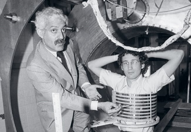

The Indomitable, as Damadian called it, was a nightmare to maintain. For one thing, it kept leaking the very-expensive helium, costing him $2,000 a week and reducing the magnet’s strength.
“In principle, the MRI machines of today are much the same thing,” says KV Ramanathan, a professor with the NMR Research Centre at the Indian Institute of Science in Bengaluru. “But advancements in the field have long since fixed the helium leak and built much-more-powerful magnets.”
Peter Mansfield, a British physicist, figured out how to speed up the scan from the initial five hours for one image, by collecting radio signals from different parts of the body at the same time.
Lauterbur and Mansfield jointly won the Nobel Prize in Medicine in 2003, for their work in this field. Damadian was the forgotten hero; of all the Nobels awarded in this chain, he never got one.
“Dr Damadian’s contribution was to apply [the NMR technique] to living tissues. That’s the big step and that’s not easy,” Nicolaas Bloembergen, winner of the 1981 Nobel Prize in Physics for his contribution to the development of laser spectroscopy, would later say. “In my opinion, Dr Raymond Damadian’s contribution is as important and as significant as the second step made by Lauterbur.”
Out in the world of medicine, the evolution of the machine continued. Amid advances in cooling systems and superconducting alloys, it has gone from a giant, steely contraption with a cage for the torso; to bulky but pristine white-and-blue tunnels closed at one end; to sleek, slim tunnels open on both sides.
The one thing that has not changed: the large, donut shaped hull and 60-cm bore, big enough for the average human body to pass through.
MRI scans have become clearer too, with better resolution and higher contrast. Contrast dyes are used for a better view of blood supply to certain vessels, organs and tumours. Functional MRI or fMRI is used to map small changes in brain activity.
“We have had immense advances in the field since it was invented, but our advances in general keep making the technology more expensive,” says Vikas Gulani, Fred Jenner Hodges professor and chair at the department of radiology at the University of Michigan. The magnets that drive the machines are expensive; the cooling systems depend on rare liquid helium.
New machines are overcoming some of these hurdles. (Click here for more on India’s own innovations in this field.)
They’re getting smaller too, and this could reduce cost (though it could also impact image quality). In 2020, for instance, the US Food and Drug Administration approved the world’s first bedside low-field MRI scanner on wheels, for brain scans. “Such efforts are starting to address the problem of access creatively,” Gulani says.
***
Quietly, in the background, MRI and the technology that first enabled it — NMR spectroscopy — are meanwhile infiltrating areas far beyond medicine. These include archaeology, chemical analysis, geology, botany and food sciences.
NMR is being used to estimate oil and gas shale deposits at various depths, and help detect the right spots from which to initiate drilling and fracking. It is being tested for its potential to assess groundwater content and monitor groundwater reserves.
NMR spectroscopy is used to analyse the physical and chemical properties (and therefore strengths and composition) of polymers, plastics and rubbers, piping, paints and tyres, in industrial plants. In the pharmaceutical industry, it is used in the research, development and design of new drugs, to identify structures of synthesised molecules, impurities, and the effects of drugs on target organs.
Meanwhile, in the field of botany, scientists at the University of Western Sydney have used high-resolution MRI to trace how water is transported, via microscopic vessels, inside living plants, in the hope that new findings could help scientists engineer sturdier, more-resilient crops amid the climate crisis. MRIs have also been used to observe how embolisms (or fatal air bubbles) form in plant species, blocking the passage of water and nutrients.
Interestingly, in ancient mummies, MRIs have proven to be less useful than CT scans, because they do not have water content to leverage. Still, such scans have helped confirm CT scan findings, allowing archaeologists to estimate what might have caused a long-gone human’s death, as well as what illnesses, pains and injuries he may have navigated while alive.
In 2012, CT scans and MRIs helped scientists at the University of Zagreb in Croatia determine that a 2,900-year-old Egyptian mummy at the city’s Archaeological Museum, previously thought to be a woman, was actually a man, likely in his 20s, and who likely had diabetes.
Mummies and humans alike must, of course, be divested of all jewellery before entering the room. The magnet of an MRI machine draws all nearby metal to it so violently, that it will pass through anything that stands in its way. This is why, for many years, people with stents in their hearts or hip replacements could not undergo such a scan. Advances in medicine have since made non-metal substitutes widespread, to ensure that most patients can safely glide into the whomping magnetic machines today. If they can afford it, that is.


