E-Paper



A mini-universe: What scientists found in 1 cubic mm of brain tissue

 Prefer HTon Google


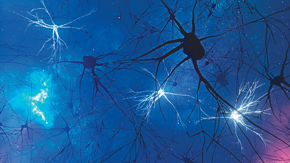
The most detailed map of a brain fragment yet threw up a range of surprises: more synapses per neuron than thought, and a lot more ‘housekeeping’ cells.
Exactly what’s going on up there?
The first-ever map of a 1-cubic-mm fragment of human brain has been completed, after 10 years, and the findings have thrown up numerous surprises.
Keep in mind, as you read, that 1 cubic mm is about half the size of a grain of rice.
The fragment, taken from the temporal cortex of an anonymous person with severe epilepsy, was analysed by researchers at the Lichtman Lab at Harvard University, with help from neuro-connectomics scholars at Google Research.
Inside, the researchers say, they found a mini-universe: 150 million synapses and 9 inches of blood vessels in a delicate tapestry, in addition to what amounts to 1,400 TB of data — proof that the brain’s wiring is far more complicated and more organised than previously thought.
The findings surprised even Jeff Lichtman, who has been studying the brain for 20 years, as head of the Lichtman lab and dean of science at Harvard University.
Among the novel findings is one that shows neuron connections aren’t all single- or two-point ones, as was previously believed. In some cases, up to 50 synapses help pass along chemical impulses.
These connections are so unexpectedly strong, it’s as if two houses, which ideally should have a single phone line connecting them, turned out to have 50 phone lines linking them instead, Lichtman has said. What’s going on here? Why are some neurons so strongly connected?
One hypothesis suggests that the super-strong links could be used to encode reflexes or other responses that need to be activated with very high reliability and speed, says Viren Jain, a senior scholar at Google Research and co-author of the study on these findings that was recently published in the journal Science.
Also found in the fragment was a surprising amount of housekeeping support in the brain.
It turns out that every neuron (or “thinking cell”) has not one but two attendant glia (or “support cells”) nearby, to act as a sort of maintenance crew. They “clean up” by regulating the chemical environment, helping repair damaged nerve tissue and removing cellular debris and pathogens, so that the neurons can function efficiently.
Shades of grey
In terms of how the fragment was studied, it was first sliced thin by a diamond-edged blade, then scanned by an electron microscope, with the resultant super-high-resolution images computationally stitched together to build a 3D map of neurons. AI-led Google tools were then used to colour-code and reconstruct the complex wiring of the brain tissue.
The eventual aim, of course, is to build such a map of a complete human brain, so that we may better understand how it works — how we work — and better understand mental illness and clinical diseases.
Such an effort would yield exabytes of data, says Jain. (An exabyte is a million terabytes, but then a brain is about 1.2 million cubic mm).
It’s anyone’s guess how many years it would take, given that we now have AI to help, but also given that we have been trying for over 2,000 years.
It was in Alexandria, in the 3rd century BCE, that neuroanatomists Herophilus and Erasistratus distinguished between the cerebrum and cerebellum, and provided what is generally considered the first scientific description of the parts of a human brain.
By the 16th century, amid the Renaissance and a renewed interest in science, dissections of humans and animals were conducted before large audiences of scholars, with some open to the general public too.
It would be centuries before the world’s first brain map was created, in 1986, representing the 302 neurons of a roundworm. “It took 16 years to create it from cross-sectional microscope images of the worm,” Jain notes. His team has lately been mapping the brains of a fruit fly, a zebra finch, and zebrafish larvae, he adds.
The results of the 1-cubic-mm study, meanwhile, have now been turned into an open-access online resource called Neuroglancer, so that scientists around the world can view findings, explore scans and run their own analyses on them.
Seeing the complexity of this tiny fragment of the human brain has been humbling, Jain says. “Even this was only possible because of AI. Indeed, we have a long way to go.”