
 E-Paper



How to scan your dragon: MRIs for non-humans drive a brain atlas programme

 Prefer HTon Google


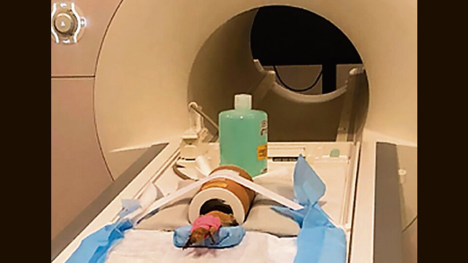
Dogs, cats, horses and sheep have had their heads scanned. So has the bearded dragon. Insects are being explored too. See why, how and what’s next.
How differently do animals’ brains work? How do they register pleasure and pain? What areas light up as they sleep, and why?
Scientists have been using magnetic resonance imaging (MRI) to try and answer such questions, creating animal brain atlases of different species.
These atlases, for now, map the brains of healthy specimens and cadavers, to establish scanning protocols and a clear view of a baseline. The aim is for these maps to eventually aid in research, and help diagnose and treat certain conditions and diseases.
Brain atlases have been created for a range of species so far, including domestic dogs, cats, horses and sheep. The most that has been learnt from these, so far, is that the balance between anaesthetising an animal safely and keeping them still enough for a scan is a science all its own.
The scans themselves are starting to identify areas of functional activation — such as what part of the brain lights up when a tail is wagged or a smack received, in cases where live specimens are studied. Deeper analysis, including details of pathologies and resting states, will be among the next steps.
Meanwhile, in one particularly unusual effort in this field, a research project in the US mapped the brain of, of all things, the chameleon-like reptile known as the central bearded dragon.
Why? Well, the Pogona vitticeps, a native of Australia, is fast becoming a pet of choice for Americans, because of its social, friendly, gentle temperament. It is one of the most common lizard species that veterinarians in the US have to work with. And it offered an intriguing departure from the canines, felines and equines that currently dominate this field.
Researchers from the University of Illinois’s Beckman Institute for Advanced Science and Technology and its College of Veterinary Medicine accordingly mapped the brain of a live bearded dragon. The areas relating to balance, movement and sight were well-developed in this species, the researchers stated, in a paper published in the journal Frontiers in Veterinary Science in May 2022.
Other reptiles with their own MRI-based brain atlases include the tokay gecko, green anole, garter snake, and tawny dragon. But because of the risks of anaesthetisation, many of these were made using cadavers rather than live specimens. “Our goal for this study was to not only provide clinicians with an anatomic reference of the bearded dragon brain, but to also establish a safe and efficient MRI and sedation protocol,” Kari Foss of the College of Veterinary Medicine, one of the authors of the study, said in a statement.
Elsewhere, scientists are studying insect brains in a similar manner, using high-resolution magnetic resonance microscopy. What’s going on in the head of a cockroach? How does a caterpillar’s grey matter differ from that of the eventual butterfly? These are questions we at Wknd are now pondering. We’ll keep you posted, amid advances in this buzzing field.


